Radiology
X-rays
Radiographs (X-rays) offer vets a non-invasive way to look at what is happening on the inside of your pet. The two main types of tissues we look at using X-rays are bones and soft tissue structures.
Conditions affecting the bones that may be diagnosed using X-rays include:
- fractures
- osteoarthritis
- spinal problems
- dislocations
- bone cancer
They can also be used to monitor healing fractures, to ensure orthopaedic surgeries have been successful, or to monitor bone growth in young animals.
Conditions affecting soft tissue structures that can be monitored using X-rays include:
- heart enlargement
- chest or lung conditions such as pneumonia or asthma
- cancer
- stomach or intestinal foreign bodies
- kidney disease
- bladder stones
- constipation
X-rays are also used in dentistry to assess what is happening to the roots of teeth underneath the gum line. This helps us to better diagnose dental issues in your pet that would likely be causing them pain or discomfort.
X-rays are safe for your pet and are normally conducted while they are under a light sedative to calm their nerves. Some X-rays require animals to lay in uncomfortable positions, like hip X-rays or after a fracture. For these, a general anaesthetic might be used.
What happens to my pet when they are booked in for an X-ray?
Most of our patients are admitted into hospital for the whole day to have routine X-rays taken (except in emergencies). We ask that you don’t feed your pet on the morning of their X-ray in case they require any sedatives or anaesthetics. Please click here for more information on pre-surgical care.
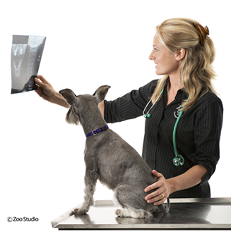
Greencross Vets clinics are equipped with high-quality radiograph equipment including X-ray machines, automatic processors, and X-ray view equipment. Once the X-rays have been processed, your veterinarian will show you the images and discuss your pet’s diagnosis and treatment plan in a discharge appointment.

 Greencross Vets


